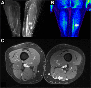
PET/MR imaging of patient with antisynthetase syndrome, a rare rheumatologic syndrome that is associated with interstitial lung disease, dermatomyositis, and polymyositis.
Researchers examine the use of combined PET/MR in their study published in the Journal of Nuclear Medicine run by the Society of Nuclear Medicine and Molecular Imaging (SNMMI). With breakthroughs in finding new detectors to replace photomultiplier tubes (PMT) in traditional PET, such as avalanche photodiodes (APD), Geiger-mode APDs (AKA silicon PMTs or solid-state PMTs), and silicon photomultipliers (SiPM), tolerance to magnetic fields and time-of-flight PET scans became possible. X-Z LAB’s Basic Detector Module (BDM) | PET Detector Module utilizes this technology and enhances it with our patented multi-voltage threshold (MVT) algorithm.
Although the researchers do not see combined PET/MR replacing PET/CT for clinical diagnostics, they believe it will likely overtake PET/CT in small-animal and clinical research. MR does not require additional ionizing radiation like CT does, and it provides much better soft-tissue contrast as well. Its low sensitivity is compensated for with PET, which has high sensitivity, and MR makes up for PET’s lack of anatomic landmarks for metabolic information.
Not only do they compare combined PET/MR with PET/CT, they also show the pros and cons of performing combined PET/MR versus sequential PET/MR. Sequential PET/MR has fewer technologic challenges and allow for each modality to be used separately, which could reduce initial investment and operation costs. However, combined PET/MR has the advantage of simultaneity, acquiring multiple functional and physiologic parameters at the same time, such as temperature, blood pressure, and heart rate. This is especially important when imaging certain parts of the body, such as the abdominal area, where differences in patient movement and position makes fusing sequential images troublesome.
Overall, the authors believe that combined PET/MR will quickly become a new standard in preclinical and clinical research. In order for that to happen, though, users must undergo thorough training and protocols must be established.
Abstract
The combination of PET and MR imaging forms a powerful new imaging modality, PET/MR. The major advantages of concurrent PET/MR acquisitions range from patient comfort and increased throughput to multiparametric imaging and are evaluated and reviewed in this paper specifically with respect to their applications in research and diagnostics. Alongside the use of PET/MR in the field of preclinical research, this paper illuminates the impact of this new modality in the clinical field in such areas as neurology, oncology, and cardiology. Now that PET/MR technology has matured, attention is needed on standardizing education for nuclear and radiologic technologists and physicians specifically for this combined modality. Furthermore, the impact of this combined modality on health economy needs to be addressed in more detail to further propel its use.
Document Credits
Hans F. Wehrl1, Alexander W. Sauter1,2, Mathew R. Divine1 and Bernd J. Pichler1
1Werner Siemens Imaging Center, Department for Preclinical Imaging and Radiopharmacy, Eberhard Karls University Tuebingen, Tuebingen, Germany
2Department of Diagnostic and Interventional Radiology, Eberhard Karls University Tuebingen, Tuebingen, Germany
Wehrl HF, Sauter AW, Divine MR, Pichler BJ. 2015. Combined PET/MR: A Technology Becomes Mature. J Nucl Med. 56(2):165-168.
